Rugal Folds Dog Endoscopy Essentials Upper Gastrointestinal Endoscopy Techniques Part
Scanning the ventral abdomen of the standing dog or cat may allow improved visualization of the dependent pylorus and gastric body. Nothing adds character like a wrinkled face. Gastric rugae can be recognized in the fundus and body of the stomach with the visibility and thickness.
Radiology Case of the Week Canine Gastrointestinal Foreign Body
Gastric biopsy specimens showed histologic evidence of mild erosive subacute gastritis. The rugal folds may be identified if the stomach is relatively empty but disappear when it is distended. In dogs, the cervical esophagus has longitudinal mucosal folds that disappear when the lumen is fully insufflated.
Ric inflammation it is important to note that the.
However, right and left lateral recumbency may assist. Without fluid distention the dorsal aspect is often not visualized because of the presence of. Longitudinal mucosal folds, which disappear when the lumen is fully insufflated, are encountered in the canine cervical esophagus. Anatomically, the ileum is contiguous to the ileocecocolic junction in the cat, leading to the ascending colon.
These thickness measurements are taken in between rugal folds. In both species, the outline of the trachea in the ventral wall. They add character, personality, and a certain je ne sais quoi that makes them utterly irresistible. In both species, note the outline of the:

Radiology Case of the Week Canine Gastrointestinal Foreign Body
These adorable folds and creases are more than just skin deep;
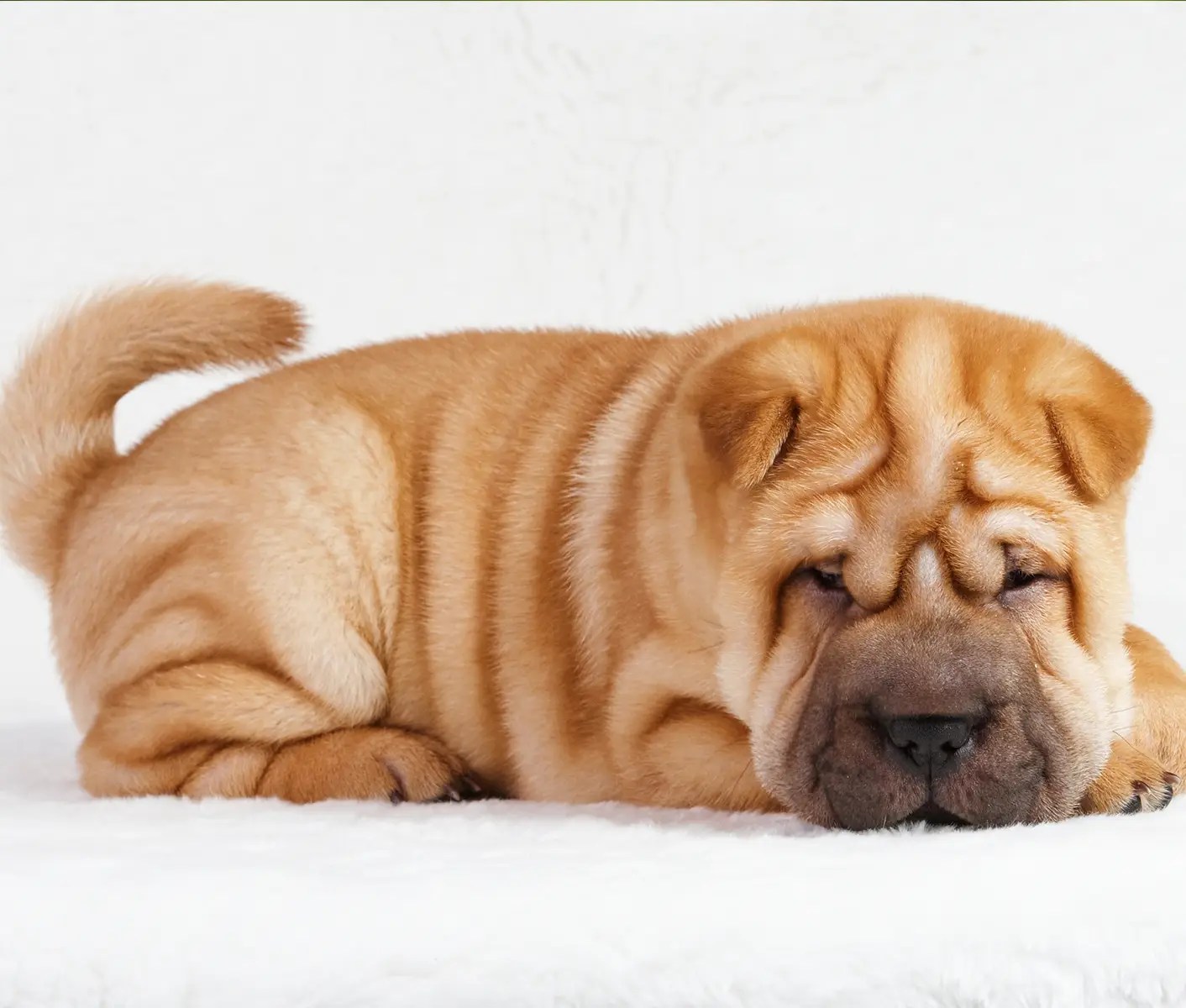
Reference range of normal gastric mucosal fold thickness, 1 to 8 mm, was defined by this study for dogs of any breed weighing between 2 and 50 kg. These are actually called “ruga” or “rugal folds” and play a crucial role in a dog’s lip anatomy. Trachea in the ventral wall of the. Ileum of dogs has a prominent submucosa (figure 4).
Histiocytic ulcerative colitis was also identified. One hundred two dogs without known. These folds are a natural part of a dog’s anatomy and can vary in size and prominence depending on the breed. Some dogs have the perfect combination of adorability and uniqueness;

Gama Bull Kennel on Instagram “JK RUGAL 🌈 🦾 completo de DNA e
Ruga or rugal folds are the wrinkles or folds on a dog’s lips.
The stomach is recognized by its location caudal to the liver and by the presence of rugal folds. Whether they have deep folds from birth or develop them with. The primary purpose of these folds is to help your furry friend grip onto their food while they chew,. Al feline rugal folds (a).
Animals are usually scanned in dorsal recumbency; Fluid and gas may be seen swirling around in the stomach as a result of peristalsis.

Foreign Body Ingestion Can Be LifeThreatening Snodgrass Veterinary

Endoscopy Essentials Upper Gastrointestinal Endoscopy Techniques, Part
What Is Skin Fold Dermatitis In Dogs
